Imaging early human brain development
Speaker
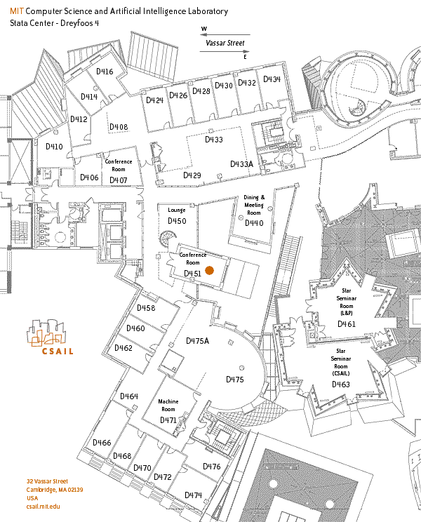
Ali Gholipour
Boston Children's Hospital, Harvard Medical School
Host
Polina Golland
MIT CSAIL
The human brain undergoes its most rapid formative growth during the
fetal period, in which a sequence of amazingly programmed processes
eventually forms the most complex living organ known. Until recently,
our ability to study brain development in-utero was limited to crude
linear measurements of the brain anatomy on prenatal ultrasound or
fetal MRI slices. With the advent of motion-corrected, robust
super-resolution MRI reconstruction, the field progressed rapidly with
new tools and resources such as atlases that have enabled mapping and
analyzing the development of the brain microstructure and function
before birth. These technological advances in fetal imaging are
crucial to study mechanisms and patterns of normal and altered brain
development. In this talk, we will review the technical advances that
have made the foundation of next-generation in-vivo fetal neuroimaging
techniques. We will discuss motion-robust diffusion-weighted MRI that
offers a unique ability to study the development of the fetal brain
connectome. In addition to slice-to-volume reconstruction, atlas
construction, and their applications, we will discuss how deep
learning techniques have contributed to advancing various fetal MRI
technologies at the acquisition and the post-acquisition processing
steps.